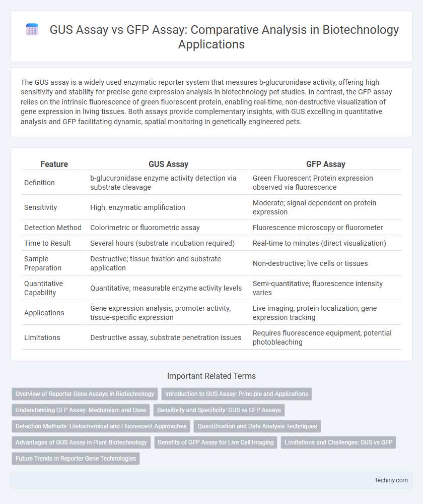
The GUS assay is a widely used enzymatic reporter system that measures b-glucuronidase activity, offering high sensitivity and stability for precise gene expression analysis in biotechnology pet studies. In contrast, the GFP assay relies on the intrinsic fluorescence of green fluorescent protein, enabling real-time, non-destructive visualization of gene expression in living tissues. Both assays provide complementary insights, with GUS excelling in quantitative analysis and GFP facilitating dynamic, spatial monitoring in genetically engineered pets.
Table of Comparison
| Feature | GUS Assay | GFP Assay |
|---|---|---|
| Definition | b-glucuronidase enzyme activity detection via substrate cleavage | Green Fluorescent Protein expression observed via fluorescence |
| Sensitivity | High; enzymatic amplification | Moderate; signal dependent on protein expression |
| Detection Method | Colorimetric or fluorometric assay | Fluorescence microscopy or fluorometer |
| Time to Result | Several hours (substrate incubation required) | Real-time to minutes (direct visualization) |
| Sample Preparation | Destructive; tissue fixation and substrate application | Non-destructive; live cells or tissues |
| Quantitative Capability | Quantitative; measurable enzyme activity levels | Semi-quantitative; fluorescence intensity varies |
| Applications | Gene expression analysis, promoter activity, tissue-specific expression | Live imaging, protein localization, gene expression tracking |
| Limitations | Destructive assay, substrate penetration issues | Requires fluorescence equipment, potential photobleaching |
Overview of Reporter Gene Assays in Biotechnology
Reporter gene assays are essential tools in biotechnology for monitoring gene expression and cellular events, with GUS assay and GFP assay being prominent examples. GUS assay detects b-glucuronidase enzyme activity by producing a blue color change, allowing quantitative analysis through substrate conversion, while GFP assay utilizes green fluorescent protein to enable real-time, non-destructive visualization of gene expression in living cells. These assays provide complementary advantages: GUS offers high sensitivity and histochemical localization, whereas GFP facilitates dynamic monitoring without requiring cell disruption, enhancing functional genomics and genetic engineering studies.
Introduction to GUS Assay: Principle and Applications
The GUS assay utilizes the enzyme b-glucuronidase as a reporter to monitor gene expression by hydrolyzing colorless substrates into a detectable blue product, enabling precise spatial and temporal analysis in plant biotechnology. This method offers high sensitivity and stability, making it valuable for studying promoter activity, gene regulation, and transformation efficiency. Applications extend to transgenic plant research, functional genomics, and validating genetic constructs in various biological systems.
Understanding GFP Assay: Mechanism and Uses
The GFP assay utilizes the green fluorescent protein, which emits bright green fluorescence when exposed to ultraviolet or blue light, enabling visualization of gene expression in living cells. This assay offers real-time monitoring of protein localization and dynamics without cell destruction, making it ideal for studying gene regulation and cellular processes. GFP's non-invasive nature and high sensitivity provide advantages over traditional assays like the GUS assay, which requires cell lysis and substrate addition for enzymatic activity detection.
Sensitivity and Specificity: GUS vs GFP Assays
GUS assays exhibit high sensitivity due to enzymatic amplification, allowing detection of low gene expression levels, while GFP assays rely on direct fluorescence, which may be less sensitive in detecting weak signals. Specificity in GUS assays can be affected by endogenous plant b-glucuronidase activity, potentially causing background noise, whereas GFP assays offer higher specificity with minimal background in most organisms. The choice between GUS and GFP assays depends on the required sensitivity and specificity for gene expression analysis in a given experimental context.
Detection Methods: Histochemical and Fluorescent Approaches
The GUS assay utilizes histochemical staining to detect b-glucuronidase activity, resulting in a blue precipitate visible under a light microscope, making it ideal for spatial localization in tissue samples. In contrast, the GFP assay employs fluorescent detection, where green fluorescence emitted by the GFP protein is observed using fluorescence microscopy, enabling real-time monitoring and quantitative analysis of gene expression in living cells. Both methods provide complementary insights, with GUS offering precise tissue-specific visualization and GFP allowing dynamic observation of gene activity.
Quantification and Data Analysis Techniques
GUS assay provides quantitative data through spectrophotometric measurement of enzymatic activity, enabling precise analysis of gene expression levels. GFP assay allows real-time visualization and quantification using fluorescence microscopy and flow cytometry, facilitating dynamic study of protein localization and expression in living cells. Data analysis for both assays relies on standardized controls and statistical methods to ensure accuracy and reproducibility in gene expression quantification.
Advantages of GUS Assay in Plant Biotechnology
GUS assay offers high sensitivity and quantitative precision, enabling detailed analysis of gene expression patterns in plant tissues. Its stability under diverse experimental conditions facilitates long-term studies without rapid signal degradation common in fluorescent markers. The assay's cost-effectiveness and straightforward protocol make it a preferred choice for high-throughput screening in plant biotechnology research.
Benefits of GFP Assay for Live Cell Imaging
GFP assay offers significant advantages for live cell imaging due to its ability to provide real-time visualization of cellular processes without the need for substrate addition, unlike GUS assay which requires destructive sample preparation. GFP's intrinsic fluorescence enables non-invasive tracking of protein localization and dynamics within living cells, facilitating detailed temporal studies. This property enhances experimental efficiency and accuracy in monitoring gene expression and protein interactions in live biological systems.
Limitations and Challenges: GUS vs GFP
GUS assays face limitations such as the requirement for destructive tissue sampling and the inability to provide real-time monitoring of gene expression, which restricts their application in dynamic studies. GFP assays enable live-cell imaging and temporal analysis but encounter challenges including photobleaching, autofluorescence interference, and potential cytotoxicity affecting cellular physiology. Both assays require careful optimization of experimental conditions to balance sensitivity and specificity while minimizing artifacts in gene reporter analysis.
Future Trends in Reporter Gene Technologies
Emerging reporter gene technologies in biotechnology are advancing beyond traditional GUS and GFP assays, emphasizing enhanced sensitivity, real-time visualization, and multiplexing capabilities. Innovations include synthetic reporters with improved signal-to-noise ratios and non-invasive monitoring in living organisms. Integration with CRISPR-based systems and high-throughput screening platforms is set to revolutionize gene expression analysis and functional genomics.
GUS assay vs GFP assay Infographic